Correlation of levator ani muscle strength measurement between Modified Oxford Grading Scale and perineometer on pelvic organ prolapse patient

Submitted: 28 February 2021
Accepted: 15 June 2021
Published: 30 June 2021
Abstract Views: 2443
PDF: 230
Publisher's note
All claims expressed in this article are solely those of the authors and do not necessarily represent those of their affiliated organizations, or those of the publisher, the editors and the reviewers. Any product that may be evaluated in this article or claim that may be made by its manufacturer is not guaranteed or endorsed by the publisher.

Authors

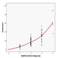
Pelvic Organ Prolapse (POP) is a debilitating condition affecting about half of all women aged of more than 60 years globally. Reduced levator ani muscle strength in POP is associated with worse symptoms and prognosis. Measurement of levator ani muscle strength can be done with several tools such as perineometer and digital palpation. However, there is currently no study regarding conformity between tests. The aim of this study is to determine the correlation between tests in POP patients. An analytic observational study using cross sectional design was done to determine conformity between perineometer and digital examination using Modified Oxford Grading Scale (MOS) in Dr Cipto Mangunkusumo National General Hospital, Indonesia during the period of July, 2018 to June, 2020. Correlation between tests was determined using Spearman test. Cut-off of perineometer reading for each MOS score was also determined. A total of 110 subjects examined with both perineometer and digital palpation were recruited to the study. Positive correlation was observed between perineometer reading and Modified Oxford Grading Scale (r = 0.790, p < 0.001). According to the result, values between 0.01 – 9.64 cmH2O correspond to very weak pressure (MOS 1); 9.65 – 22.49 cmH2O represent weak pressure (MOS 2); 22.5 – 35.24 cmH2O represent moderate pressure (MOS 3); ≥ 35.25 cmH2O represent good pressure (MOS 4). There was a strong correlation between MOS and perineometer result for measuring levator ani strength in POP patients.

Dimensions

Altmetric

PlumX Metrics

Downloads

Download data is not yet available.

Citations

Wilkins MF, Wu JM. Epidemiology of pelvic organ prolapse. Current Obst Gynecol Rep 2016;5:119-23. DOI: https://doi.org/10.1007/s13669-016-0149-z
Shah AD, Kohli N, Rajan SS, Hoyte L. The age distribution, rates, and types of surgery for pelvic organ prolapse in the USA. Int Urogynecol J 2008;19:421-8. DOI: https://doi.org/10.1007/s00192-007-0457-y
DeLancey JO. What’s new in the functional anatomy of pelvic organ prolapse? Current Opinion Obstet Gynecol 2016;28:420. DOI: https://doi.org/10.1097/GCO.0000000000000312
Weintraub AY, Glinter H, Marcus-Braun N. Narrative review of the epidemiology, diagnosis and pathophysiology of pelvic organ prolapse. Int Braz J Urol 2020;46:5-14. DOI: https://doi.org/10.1590/s1677-5538.ibju.2018.0581
Van Geelen H, Ostergard D, Sand P. A review of the impact of pregnancy and childbirth on pelvic floor function as assessed by objective measurement techniques. Int Urogynecol J 2018;29:327-38. DOI: https://doi.org/10.1007/s00192-017-3540-z
Yang X, Zhu L, Li W, et al. Comparisons of electromyography and digital palpation measurement of pelvic floor muscle strength in postpartum women with stress urinary incontinence and asymptomatic parturients: a cross-sectional study. Gynecol Obstet Invest 2019;84:599-605. DOI: https://doi.org/10.1159/000501825
Da Roza T, Mascarenhas T, Araujo M, et al. Oxford Grading Scale vs manometer for assessment of pelvic floor strength in nulliparous sports students. Physiother 2013;99:207-11. DOI: https://doi.org/10.1016/j.physio.2012.05.014
American College of Obstetricians and Gynecologists. Pelvic organ prolapse. Female Pelvic Med Reconstruct Surg 2019;25:397-408. DOI: https://doi.org/10.1097/SPV.0000000000000794
Bo K, Sherburn M. Evaluation of female pelvic-floor muscle function and strength. Phys Ther 2005;85:269-82. DOI: https://doi.org/10.1093/ptj/85.3.269
Volløyhaug I, Mørkved S, Salvesen Ø, Salvesen KÅ. Assessment of pelvic floor muscle contraction with palpation, perineometry and transperineal ultrasound: a cross‐sectional study. Ultrasound Obstet Gynecol 2016;47:768-73. DOI: https://doi.org/10.1002/uog.15731
Angelo PH, Varella LRD, de Oliveira MCE, et al. A manometry classification to assess pelvic floor muscle function in women. PLoS One 2017;12:e0187045. DOI: https://doi.org/10.1371/journal.pone.0187045
Dietz HP, Shek C, De Leon J, Steensma AB. Ballooning of the levator hiatus. Ultrasound Obstet Gynecol 2008;31:676-80. DOI: https://doi.org/10.1002/uog.5355

How to Cite

Moegni, F., & Wengkang , I. F. O. (2021). Correlation of levator ani muscle strength measurement between Modified Oxford Grading Scale and perineometer on pelvic organ prolapse patient. Urogynaecologia, 33(1). https://doi.org/10.4081/uij.2021.268